Visual field
FDT – (Frequency-doubling technology) was developed as a fast, effective way to detect visual field loss. The FDT is the ideal visual field screening device because it only 40 seconds per eye. This quick and easy test is preformed on all patients during all comprehensive eye exams.
The Humphrey Threshold Visual Field is a special automated procedure used to perform perimetry, a test that measures the entire area of peripheral vision that can be seen while the eye is focused on a central point. During this test, lights of varying intensities appear in different parts of the visual field while the patient’s eye is focused on a certain spot. The perception of these lights is charted and then compared to results of a healthy eye at the same age of the patient in order to determine if any damage has occurred. This procedure is performed quickly and easily in about 15 minutes, and is effective in diagnosing and monitoring the progress of glaucoma. .
Patients with glaucoma will often undergo this test on a regular basis in order to determine how quickly the disease is progressing. The Humphrey Visual Field test can also be used to detect conditions within the optic nerve of the eye, and certain neurological conditions as well.
OCT (Optical Coherence Tomography)
OCT – (Optical coherence tomography) is an advanced technology used to produce cross-sectional images of the retina, the light-sensitive lining on the back of the eye where light rays focus to produce vision. These images can help with the detection and treatment of serious eye conditions such as retinal swelling from diabetic retinopathy, glaucoma, and age-related macular degeneration (AMD).
Optilight IPL (Intense Pulsed Light)
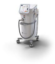
OptiLight by Lumenis is a light-based, non-invasive treatment focused on the skin around the eyes to manage dry eye. The first and only IPL FDA-approved for dry eye management. The treatment is safe, gentle, and is backed by more than 20 clinical studies.

OptiLight with Lumenis patented Optimal Pulse Technology uses precise, intense broad spectrum light to address the inflammation – one of the key underlining factors in dry eye disease due to MGD. OptiLight was specifically developed to reach the delicate contours of the treated area safely, effectively and gently. OptiLight sets new treatment standards and helps patients to regain health, control and joy. Backed by numerous clinical studies, OptiLight’s novel optimal pulse technology(OPT) provides a transformative, safe, and efficacious way to improve Dry Eye disease due to MGD.
iLux
The iLux® is used to heat and compress glands in the eyelids of adult patients with a specific type of dry eye disease called Meibomian Gland Dysfunction (MGD), also known as evaporative dry eye. It uses thermal pulsation to deliver gentle warming and pressure directly to your eyelids to unblock meibomian glands.
Fundus Photography
Fundus photography is very useful to document the natural state of the back of the eye. It is used to document and diagnose certain eye conditions. It is important to document the findings of retinal diseases and conditions, especially diabetic eye disease findings, macular degeneration, macular holes and retinal tears and detachments. We take fundus photos of all of our patients at their annual compressive exams which serves as a reference and allows us to monitor for changes in consecutive years.
Corneal Topography
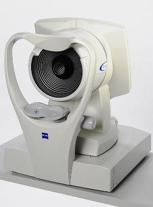
Corneal Topography is a non-invasive means of mapping the curve of the cornea’s surface. Evaluating the surface curvature of the cornea is critical in determining your quality of vision because the cornea is responsible for approximately 70% of the eye’s refractive activities. Corneal topography provides an us with a three-dimensional map of the cornea, which can assist in:
- Fitting contact lenses
- Planning LASIK or other refractive surgeries
- Screening for keratoconus and determining its severity


